Madam Butterfly
- Ask the DOC
Rockaway Stuff
- 0
- 2 minutes read

By Peter Galvin, MD
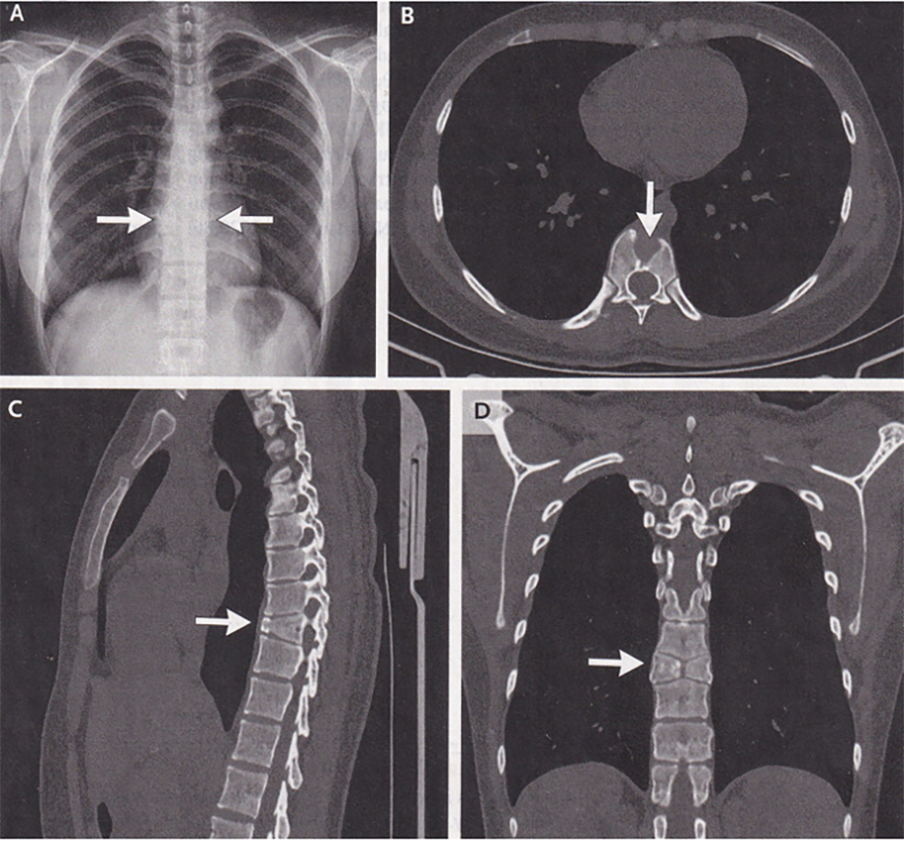
A 21-year-old woman presented to the pulmonary clinic at the Fourth Affiliated Hospital of China Medical University in Shenyang, China with a seven-day history of sore throat and cough. On routine chest X ray, she was noted to have a spinal abnormality. She denied a history of back pain. Physical examination was notable only for mild pharyngeal erythema (throat redness). The X ray showed normal lungs and two hemivertebrae (Panel A, arrows) at the ninth thoracic vertebral body (T9). A subsequent CT scan of the chest showed a sagittal cleft (Panel B, arrow). The sagittal plane runs from head to toe and divides the body into right and left sides. The CT also showed anterior wedging (Panel C, arrow) on the parasagittal view (from the side). On the coronal view (from the front), it showed symmetric, triangular hemivertebrae resembling the wings of a butterfly at T9 (Panel D, arrow). A diagnosis of butterfly vertebra was made.
A butterfly vertebra is a rare congenital (fetal) anomaly that results from a lack of fusion of the two lateral ossification (bone formation) centers during embryonic development. It is usually asymptomatic and is usually discovered incidentally on imaging, most commonly in the thoracic and lumbar spine. An incidental finding like this that is discovered during imaging for a different reason is called a red herring. A butterfly vertebra may occur in isolation, like this case, or may be associated with other congenital anomalies. A butterfly vertebra is a benign finding and does not lead to or cause any serious spinal conditions.
Reassurance regarding the benign nature of the vertebral finding was given to the patient. Supportive care was recommended for the patient’s sore throat and cough, which was attributed to a viral upper respiratory infection.
Please direct questions or comments to editor@rockawaytimes.com