Half Baked
- Ask the DOC
Rockaway Stuff
- 0
- 2 minutes read

By Peter Galvin, MD
A 63-year-old woman with psoriatic arthritis involving her knees presented to the rheumatology clinic with a 9-month history of worsening left knee pain and swelling. Her psoriatic arthritis had recently been well controlled with leflunomide and monthly golimumab injections (both are immune system suppressants). Physical examination was notable for a nontender, palpable mass in the left popliteal fossa (back side of the knee) that was more prominent when the patient was standing with her knee fully extended (Panel A). A musculoskeletal ultrasound study done in the clinic (Panel B) showed a well-defined, anechoic, fluid-filled structure resembling a cartoon “speech bubble”, with a neck extending down into the joint space between the medial head of the gastrocnemius muscle (blue outline) and the semimembranosus tendon (red outline). A diagnosis of a Baker’s cyst was made.
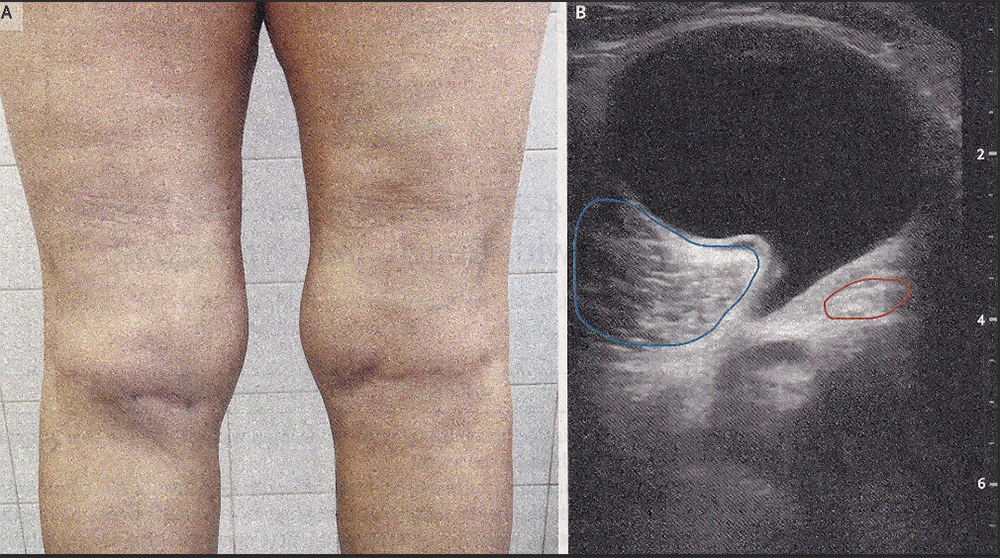
A Baker’s cyst, also known as a popliteal synovial cyst, results when synovial fluid (joint-space fluid) seeps from the knee joint and flows into and accumulates into the gastrocnemius-semimembranosus bursa (sac). Baker’s cysts are associated with underlying joint disorders including osteoarthritis, traumatic injury, or inflammatory arthritis (as in this case). Imaging studies are not always needed to make the diagnosis but may be helpful to rule out other conditions. Ultrasound-guided aspiration of the cyst was performed, and an intracystic steroid injection was given. The patient’s knee pain abated shortly after treatment, and she remained pain-free as of the two-month follow-up. She will need regular follow-ups as the fluid can reaccumulate.
Please direct questions and comments to editor@rockawaytimes.com