It Ain’t Frosty
- NEWS
Rockaway Stuff
- 0
- 3 minutes read

By Peter Galvin, MD
This case is from Wenzhou People’s Hospital in Wenzhou, China. A previously healthy 47-year-old woman presented to the neurology clinic with a one-month history of worsening headaches associated with blurry vision, galactorrhea (milk secretion from the breast in a non-pregnant woman), and irregular menstrual cycles. Physical examination was notable for reduced visual acuity of 20/50 in both eyes, with normal visual fields and eye movements. An MRI scan of the head showed an enlarged pituitary gland (gland at the base of the brain that controls many endocrine systems) protruding from the sella tursica. The sella tursica, Latin for Turkish saddle, is a saddle-shaped depression in the sphenoid bone, which comprises both part of the base of the brain and roof of the sinuses and is found in both humans and apes. Normally, the pituitary sits in the bottom of the sella tursica and is connected to the brain via an upward stalk. In this scan, the enlarged pituitary extends upward into the suprasellar cistern and is compressing the optic chiasm (where the optic nerves cross each other with the pituitary stalk in the center). This is known as the “snowman sign” (see Panel A, center).
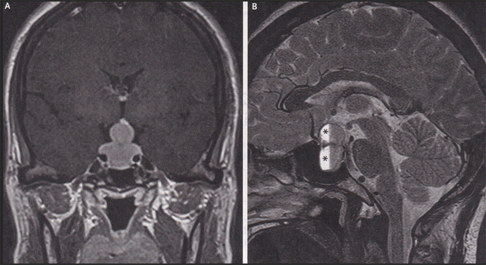
In Panel B, at asterisks, intrapituitary hemorrhage is noted. The snowman sign occurs when a slow-growing tumor that originates in the sella tursica gets indented by the roof of the sella in its upward growth. Often, although not in this case, a pituitary tumor like this will press on the optic nerves causing bitemporal hemianopsia. That is the loss of the outer side of the visual field in both eyes. It is the outer side of the visual fields because the optic nerves have already crossed each other, so signals from the outer visual fields are now on the inner side of each nerve, with the pituitary tumor in the middle. In fact, in the absence of trauma, bitemporal hemianopsia is pathognomonic (nothing else causes it) for a pituitary tumor.
The patient’s serum prolactin level (hormone made in the pituitary that stimulates breast milk production) was 200 (reference range 3.3 to 26.7 in a non-pregnant woman), and she had both adrenal and thyroid hormone deficiencies (both glands are controlled by the pituitary gland). A diagnosis of a prolactin-secreting pituitary macroadenoma was made. Transsphenoidal surgical resection was performed (removed via the nose). Her symptoms resolved on postoperative day three, and one year later a repeat MRI showed pituitary tissue in the sella tursica and no other abnormalities.
This case demonstrates the fact that while adenomas (and meningiomas) are benign growths and not malignant, there is no such thing as a “benign” brain tumor because the brain sits within the skull with no room for expansion, so anything that grows in the brain must displace normal brain tissue.
Please direct questions or comments to editor@rockawaytimes.com